ImagePro Radiology · Bone health
Bone density
Understand your bone health.
Helpful guidance before, during and after your DXA scan.
A clearer picture
of your bone health.
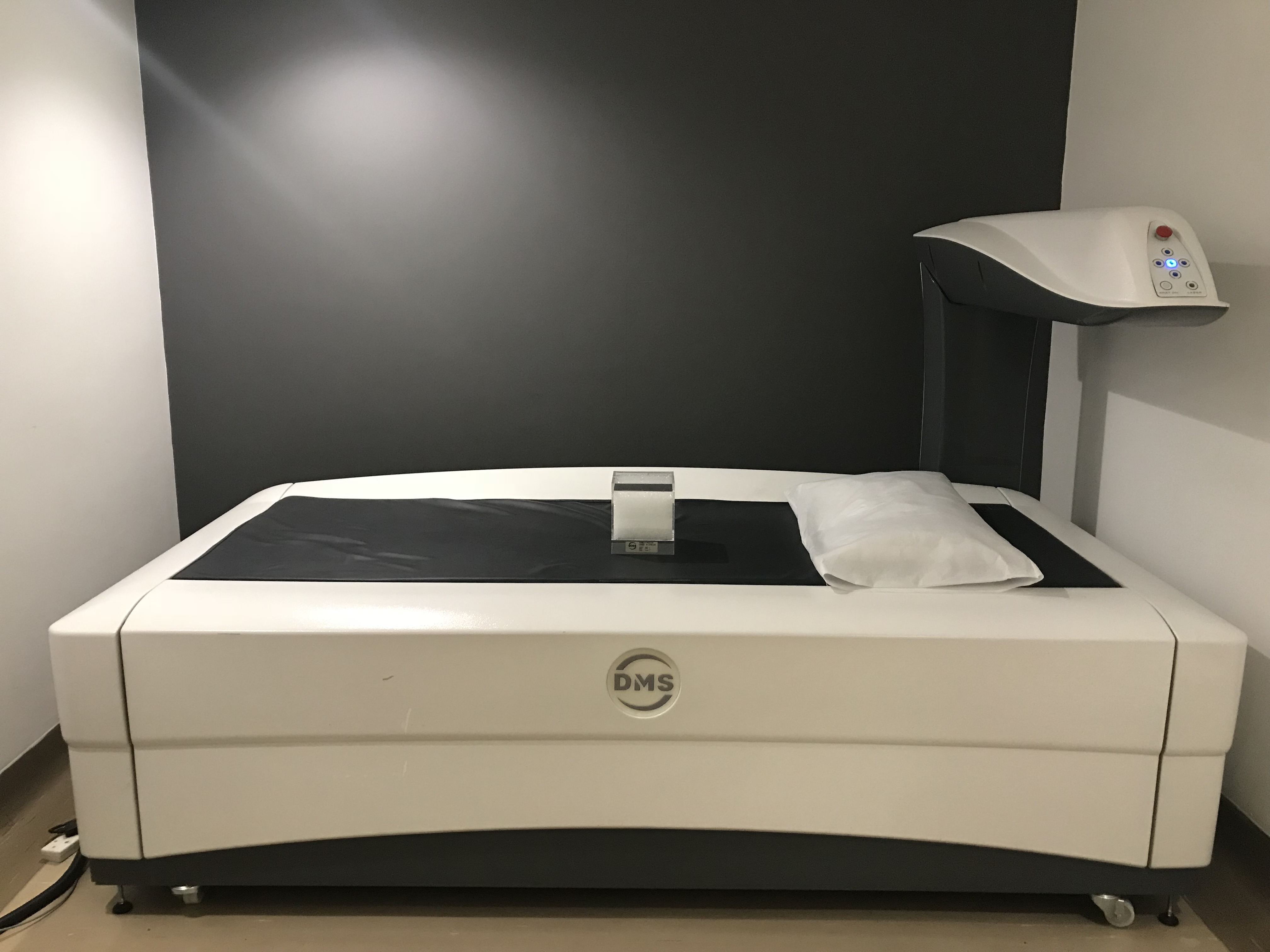
A bone-density scan measures bone mineral density. Your doctor uses the findings alongside your medical history to assess bone health and plan your care.
Also called DXA or DEXA, this examination can help diagnose osteoporosis and monitor changes over time.
Understanding bone health
What is osteoporosis?
Osteoporosis weakens bones and makes them more likely to fracture. It can develop without obvious symptoms, so a risk assessment may be useful before a fracture occurs.
It can affect both women and men. Your doctor can advise whether a bone-density scan is appropriate for you.
Individual assessment
When might a scan help?
Your doctor may consider testing because of:
- A fracture after a minor fall or injury.
- Increasing age or changes after menopause.
- A family history of osteoporosis or hip fracture.
- Long-term corticosteroid treatment or conditions associated with bone loss.
The decision and timing depend on your individual risk, rather than one starting age for everyone.
Before your visit
Preparing for your scan.
Food and calcium supplements
You can usually eat normally. Avoid calcium supplements for at least 24 hours before the scan; check with the branch if you need advice about your prescribed supplements.
Clothing and metal objects
Choose comfortable clothes without metal fasteners near the area being scanned. You may be asked to change into a gown.
Recent contrast or nuclear-medicine examinations
Tell the booking team if you recently had a barium study, a contrast-enhanced CT or a nuclear-medicine scan. These can affect measurements; the team will advise whether your appointment should be delayed.
Pregnancy or possible pregnancy
Tell the booking team and radiographer before the examination if you are pregnant or may be pregnant.
Previous scans and positioning needs
Bring your previous bone-density reports and tell us about hip replacements, spinal surgery or difficulty lying flat. This helps the team plan your scan and compare results where possible.
Your examination
What to expect.
- Check in and discuss your historyThe team confirms your referral and relevant health information.
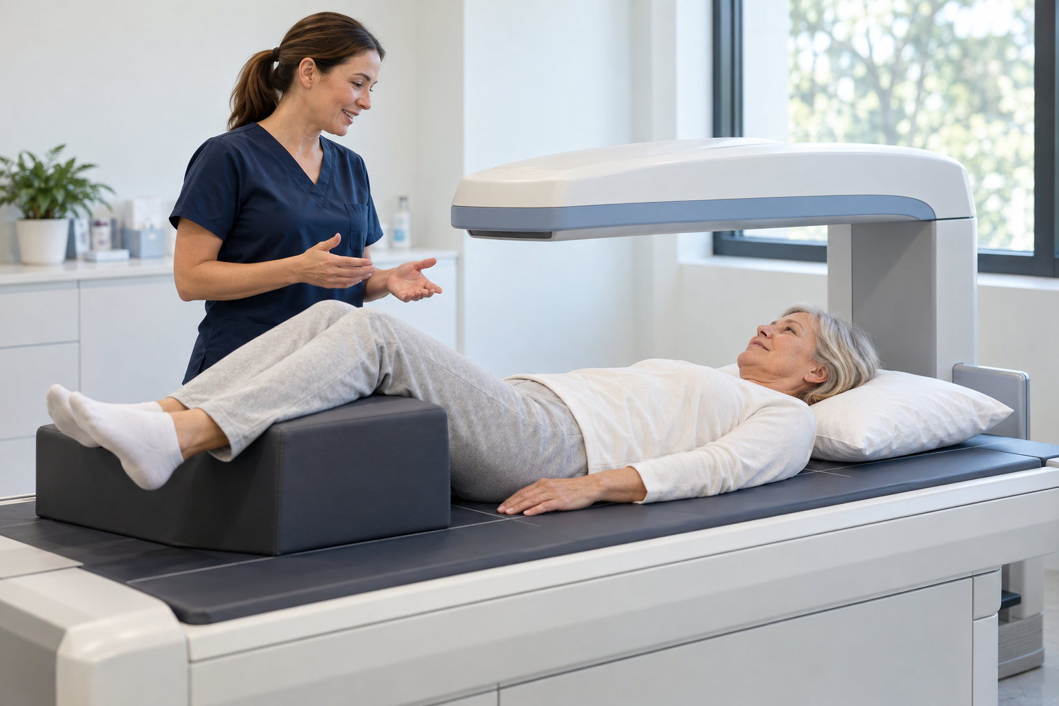
- Lie on an open examination tableThe radiographer helps position you. There is no enclosed tunnel.
- Keep still during the measurementsA scanning arm moves over the area. Measurements usually include the hip and lower spine; the forearm may be used when appropriate.
- Confirm your next stepAsk when your report will be available and discuss the findings with your referring doctor.
Allow around 20 minutes for the scan, plus check-in time. Positioning may be uncomfortable if you have pain or limited movement; let the radiographer know.
After your scan
Understanding your results.
Your report and fracture risk
Your doctor considers the measurements together with your history and other risk factors. A scan helps assess fracture risk but cannot predict with certainty whether you will break a bone.
What are T-scores and Z-scores?
A T-score compares your measurement with a young-adult reference, while a Z-score compares it with an age-matched reference. Your clinician will explain which score is relevant to you and what it means.
Will I need another scan?
Your doctor will recommend a follow-up interval based on your results, treatment and risk. Where possible, return to the same facility and scanner to support comparison.
Can I return to my usual activities?
Most people can leave and continue their normal activities after the examination. Follow any instructions the team gives you.
Speak to our team
Arrange your bone-density scan.
Contact your preferred branch to confirm bone-density availability, preparation and booking requirements. Sending an enquiry does not reserve an appointment.
Further patient information: RadiologyInfo.org: Bone density scan ↗. Follow the instructions supplied for your own examination.